What is Osteoarthritis?
Osteoarthritis is a degenerative disease of our joints that causes them to lose their supporting cartilage.
In my first post on Arthritis, at the conclusion, I had mentioned that my next topic will be on a joint disease called Osteoarthritis.
In this article, I shall be discussing what osteoarthritis is all about and what are its signs and symptoms. Let us now go back to the previous article, back to the structure of the joint.
Osteoarthritis is a degenerative joint disease involving the cartilage part of the joint. This results in the two bones of the joint grating against each other causing severe pain in movement. Sometimes there is swelling of the surrounding soft tissue like the muscles causing stiffness of the joints.
This causes-
a] pain in movement, sometimes at rest
b] a feeling of the bones grating. This can be felt like creaking, especially in the knees.
The cartilage gets worn out eventually and the joint may get deformed resulting in disability.
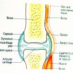
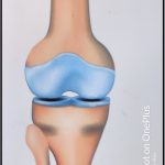
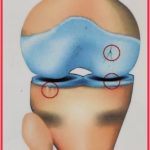
A look at the picture [click on it for a larger view] will show you that at the end part of bones facing each other there is a part called Articular Cartilage.
Table of Contents
What is Cartilage?
Cartilage is, in common man’s language, gristle. It is a tissue that:-
1] is elastic, tough and durable
2] separates two ends of the bone from directly touching each other
3] does not have a blood supply.
It gets its nutrition and oxygen from the fluid[synovial] in the joint cavity. As an example, this picture of a chicken leg below shows exactly how the cartilage looks like-
What happens when we move our joints?
Movement of the joint results in alternate compression and relaxation of the cartilage.
An increase in pressure compresses the cartilage. This squeezes out the fluid along with waste material from the cartilage.
During relaxation, the opposite occurs- fluid and nutrients get absorbed by the cartilage.
So, the health of our cartilage depends upon the regular use of our joints. In other words, regular exercise can keep your joints as well as your cartilage healthy.
So, what happens in Osteoarthritis?
As we age, changes start occurring in almost all parts of our body such as wrinkling of the skin, greying of hair, etc.
A change in the structure of our joints is also observed. This leads to the following changes-
There is a reduction of the lubricating synovial fluid leading to the grittiness of the joints.
The cartilage supporting the joint starts getting frayed and eventually disappears partially or fully.
This results in the bony ends touching each other. The bony ends of the joint contain sensory nerves. When the bones start rubbing against each other, we get pain.
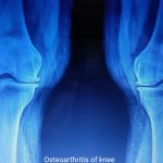
This results in a crippling sort of disability called subluxation [see the pictures below] in medical terms i.e. a part of the joint sways towards the part where the cartilage is lost.
The picture on the left-hand shows the X-ray of a normal structure of a healthy knee joint and on the right, a knee joint affected by Osteoarthritis.

Note the reduction in joint space with mild subluxation on the inner side of the affected joint.[More when we come to OA of the Knee joint]
Forms of Osteoarthritis
There are 3 forms of osteoarthritis mostly noticed in different people, notably-
1] The Fingers: –
These are visible as knotty growths in the 2nd and 3rd joints. They are painless but they alter the cosmetic appearance of the hand, as shown in this picture below-
This is the picture of a 70-year-old male patient who had undergone knee replacement for osteoarthritis 5 years back. Recently he noticed changes in the fingers of both hands in the second and third joints along with pain and stiffness.
Note the knotty appearance of the second joint of the index and middle fingers.
These swellings are also known as Heberden’s node, named after the British surgeon who first described it.
2] The Vertebral Column: –
The vertebral column is formed by a set of bones called vertebrae.
This starts from the base of the skull to the small of the back, ending in between the two buttocks.
The vertebral column is made up of –
7 vertebrae in the neck [ cervical vertebrae]
11 in the chest [ dorsal vertebrae]
5 in the trunk [lumbar vertebrae]
and 1 single large, fused vertebra called sacrum at the end of the column.
The Intervertebral Disc
In between two vertebrae, there is a disc space [as shown in the two x-ray pictures to the left and right below, of the trunk or lumbar vertebrae]. This is formed by a fibrocartilagenous structure called the intervertebral disc. Click the link below to know the anatomy of the spine or vertebral column-
In our lifetime, we move our back and neck many times every day. You can notice this when somebody is speaking- watch the number of times he or she moves their neck. These movements cause a condition called cervical spondylosis.
Normal movements like forward and backward bending, over some time, causes degeneration of this intervertebral disc, leading to a reduction in disc space. This causes compression of the spinal nerve roots giving rise to pain on movement of the neck which is termed cervical radiculopathy.
What happens when the disc is worn out?
This causes the loss of the disc space, [as shown in this x-ray picture ⇑ above] eventually causing the vertebrae to override against each other. This causes pain and formation of new bone spurs called osteophytes.
Sometimes there is a fusion of the two vertebrae causing stiffness in the neck or the lower back.
What happens next?
As the condition progresses, there is more compression of the nerve roots that come out from the spinal cord through the gaps in between two vertebrae.
This can cause tingling and numbness in the neck and the lower extremities.
All these changes put together are called Lumbar Spondylitis or Spondylosis in the back and the legs.
A similar change occurs in the vertebrae of the neck also which causes problems in the arms and parts of the chest supplied by the nerves coming out from the spinal cord in the neck. This is called Cervical spondylitis.
3] The Weight-Bearing Joints
In our body, the major joints that bear the brunt of our body weight are the hip joint, knee joint, and ankle. Because of the constant use of these joints and an increase in our body weight, there occur degenerative changes of the cartilage. These joints then lose their cartilage totally until the bones touch each other.
What happens next?
With movement, and because there is little or no cartilage, the bones grind against each other and this causes the pain of osteoarthritis. There is the formation of new bone called osteophytes. These new growths try to give support to the joint by increasing the area of the joint, which then appears as swelling of the joints. Over some time, the ends of bone touching each other become smooth and the pain lessens or disappears. This is called eburnation.
How does this look?
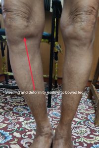
As the joint gets splayed, in the knee joint, there is movement of the bones-either to the inner or outer side of the joint. There is a bowing of the leg. The pictures shown below will give you an idea of how this deformity appears to our eyes.
This is a picture of the knee joints showing advanced osteoarthritis in an 87-year-old person.
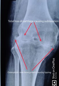
On the right is the X-ray of the same affected joint.
Note the splaying of the right knee giving rise to a bow-legged appearance. There is also swelling surrounding the joint- this is due to bursitis[inflammation of the bursa].
You can also note the new bone formation. This is called lipping, as it appears like a lip.
As the days go by, this will become more splayed, and this patient may either need the support of a cane for walking or he will have to undergo a total knee replacement operation.
You can also note the increased porosity of the bones. This condition is due to calcium deficiency and is called Osteoporosis [ brittle bone- more about this in my future article] and is a common cause of fractures of the bones in the old age.
Osteoarthritis [OA]- Signs and Symptoms
Now that we have understood different types of OA, we now proceed to learn its signs and symptoms.
Osteoarthritis progresses in stages and each stage has its own signs and symptoms. The different stages in its development can be understood by studying these diagrams of the development of OA in a knee joint shown below: –
Stage 1 OA-
There is minimal loss of cartilage. The pain in this stage is also minimal. The movement of the joint is not restricted.
Stage 2 OA-
In this stage, the first signs of wear in the cartilage can be seen. It begins to break down and cracks appear on the cartilage. The joint space starts narrowing and pain and swelling due to fluid accumulation occurs.
Here the patient may or may not complain of the stiffness of the joint in the mornings. Movements of the joint are slightly restricted.
Stage 3 OA-
In this stage, the cracks in the cartilage start expanding and the bones start touching each other. There is increased pain and more fluid accumulation. This is called effusion.
The swelling of the joint is visible [as seen in the picture knee joint of the patient shown above]. Movement of the joint is very painful, and he has to take the support of a cane to move about.
Stage 4 OA-
In this stage, the cartilage is almost totally frayed, so much so that the two opposite bones rest on each other.
There is excruciating pain on movement initially and as it becomes unbearable, the doctor may advise total knee replacement [TKR] operation.
If the patient bears the pain, after some time, the ends of the bones become smooth with the appearance of osteophytes and the pain may disappear completely.
You may have one or two types of the above-mentioned osteoarthritis at the same time.
So, you see, Osteoarthritis is quite a common condition that accompanies aging and wear and tear of the cartilage of the joint involved. This you may have observed in people you have around you like parents, grandparents or family friends.
Having said this, we will learn all about how to manage osteoarthritis in my next post. In that post, I will be discussing the investigations and treatment of osteoarthritis. So, stay tuned in for my next article.
Useful link on Osteoarthritis- Curearthritis
If you found this article useful, you can share it by clicking the quote given below-
Knowing what to expect in Osteoarthritis, one can delay its effect by taking proper care of the affected joint by its proper use, meaning reducing wear and tear. Share on X