What are the uses of getting a CT-Angiogram done?
After learning about CT angiogram , also known as computed tomography angiography cta, done by a procedure called CT-angiography, we move ahead to learn more about it, and its uses.
In this article we will learn how doing a CT-angiography done in a suspected case of coronary heart disease can help the patient to prevent future events like heart attack. A multislice ct angiography can help us learn about
So, let’s begin by-
Stages in Assessing Plaque Health in the Heart
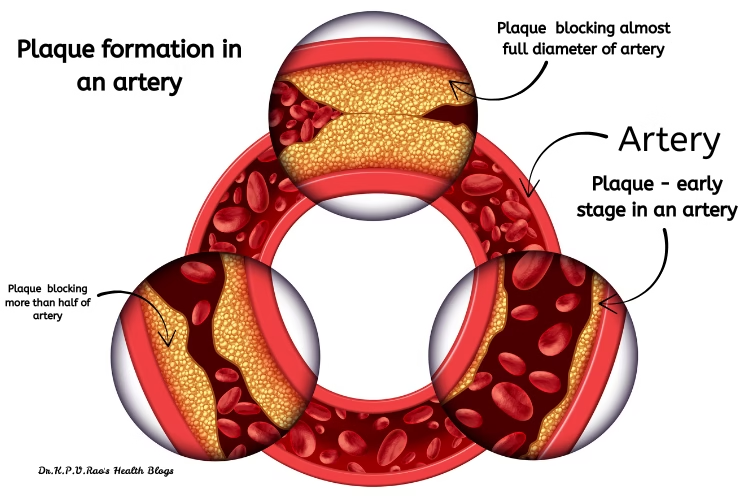
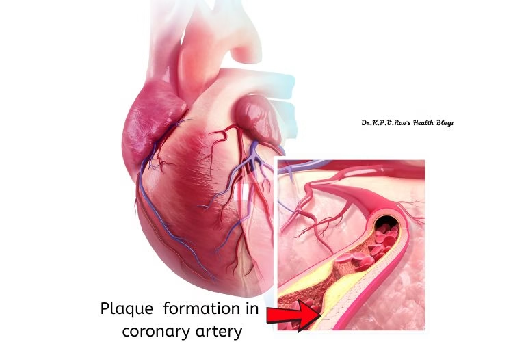
To begin with, let’s understand what a plaque means. Plaque is a deposit of cholesterol in the wall of the coronary or heart artery. Overtime, this plaque grows and then blocks the lumen of the artery, thereby cutting off the blood supply to the heart muscle causing injury or death of the muscle in that area.
The picture below shows how a small plaque initially progresses to block an artery fully:

The evaluation of plaque health within the coronary arteries is crucial for understanding cardiovascular risks and guiding treatment strategies.
This assessment is typically conducted through several distinct stages, starting with the classification of plaques, which can vary in morphology and composition.
Classification of plaques in CT-Angiography of Heart
The two primary types of plaques identified in CT-angiography of the heart are stable (calcified) and unstable (non-calcified or mixed).
Once the plaque is classified, the next stage involves visualization techniques employed by CT-angiography. Advanced imaging allows for detailed assessment of plaque characteristics, such as thickness, density, and the presence of a lipid pool.
These details are essential because they provide insights into the stability of the plaque, informing clinical decisions about the necessity for intervention or closer monitoring. High-resolution CT scans can reveal subtle changes in plaque structure, highlighting areas that may warrant further investigation or therapeutic intervention.
Plaque Calcium Score Measurements using CT-angiography
Moreover, quantitative measurements such as the Agatston score can assist cardiologists in assessing the overall burden of coronary artery disease.
This scoring method evaluates calcium levels within plaques, offering a reliable estimate of plaque severity and the potential for adverse events. The correlation between plaque health assessment via CT-angiography of the heart and patient prognosis is significant as it contributes to risk stratification and individualized treatment planning. Consequently, understanding plaque health is not merely an academic exercise but carries substantial implications for patient outcomes and long-term management of cardiovascular health.
Importance of Plaque Calcium Score in Heart Health
Plaque calcium score measurements are an important advancement in assessing the cardiovascular health of patients. Derived from CT-angiography of the heart, these measurements utilize advanced imaging techniques to detect and quantify the amount of calcified plaque within the coronary arteries. This information plays a critical role in evaluating a person’s risk for cardiovascular events, such as heart attacks and strokes, which are often associated with atherosclerosis—a condition characterized by the buildup of plaque in the arteries.

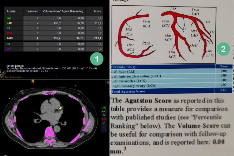
2. Image of CT- angiography of one of my patients with Agatston Score
The scoring system for calcium scoring typically employs the Agatston method, which is widely accepted in clinical practice.
In this method, the amount of calcium present in the coronary arteries is quantified and assigned a score.
The scoring ranges from zero, which indicates no detectable calcium and a low risk of heart disease, to higher scores that correlate with increased levels of calcified plaque and a corresponding rise in cardiac risk. For instance, a score of 1-10 suggests minimal risk, while scores above 400 are associated with a higher likelihood of significant coronary artery disease.
These calcium scores derived from CT-angiography provide valuable insights into the overall heart health of patients. They can help guide treatment decisions, allowing healthcare providers to develop appropriate risk mitigation strategies.
Furthermore, patients with elevated scores may be advised to engage in lifestyle modifications, such as improved diet and increased physical activity, or may be candidates for pharmacotherapy to lower their cardiovascular risk.
Understanding plaque calcium score measurements, in conjunction with other clinical assessments, is essential for effective cardiovascular risk management.
Learn more about what and how the CT-ANGIOGRAM helps in diagnosing heart disease in this informative video- CT-ANGIOGRAM
What are the benefits of CT-Angiogram
CT-angiography of the heart offers several advantages over traditional diagnostic methods such as invasive coronary angiography and other imaging modalities. One of the primary benefits of this non-invasive imaging technique is that it minimizes patient risk. Unlike traditional angiography, which requires catheter insertion into the blood vessels, CT-angiography utilizes advanced imaging technology, thereby avoiding complications associated with invasive procedures.
Another significant advantage of CT-angiography is the speed at which results are obtained. In clinical settings, timely diagnosis is crucial, especially for conditions related to coronary artery disease. A CT-angiogram can often be performed within a matter of minutes, and the imaging results are available promptly, allowing physicians to make quick and informed decisions about a patient’s care. This expedited process is beneficial for both healthcare providers and patients, as it helps in the timely management of potential heart issues.
Moreover, CT-angiography of the heart provides a detailed, three-dimensional view of the coronary arteries. This level of detail is invaluable for assessing the presence and extent of coronary artery disease, as it allows for an accurate evaluation of blood flow and any blockages that may exist within the arteries. The high-resolution images obtained can reveal not only the anatomy of the arteries but also the degree of stenosis, thus helping in tailoring appropriate treatment plans. In addition, CT-angiography can be particularly useful in patients where traditional methods might be less effective or where integrating results could enhance diagnostic accuracy.
Overall, the non-invasive nature, rapid result turnover, and detailed assessment provided by CT-angiography of the heart make it a preferred choice for many clinicians in the evaluation of cardiac conditions and patient management.
Potential Risks and Considerations
While CT-angiography of the heart is widely regarded as a valuable diagnostic tool, it is vital to consider potential risks and important factors associated with the procedure.
One primary concern is radiation exposure. CT scans utilize ionizing radiation, which can contribute to an increased risk of cancer over a lifetime, particularly in patients who undergo multiple scans.
Despite this risk, the benefits of obtaining accurate diagnostic information often outweigh the potential harm, particularly when conducted judiciously and in accordance with current clinical guidelines.
Another significant consideration involves the use of contrast agents during a CT-angiogram of the heart. Most procedures employ iodinated contrast materials, which help enhance the visibility of blood vessels.
However, these agents can provoke allergic reactions in some patients. Reactions may range from mild symptoms, such as hives or itching, to more severe responses, including anaphylaxis. Patients with known allergies to iodine or shellfish should inform their healthcare provider, who may take additional precautions or consider alternative imaging techniques if deemed necessary.
Furthermore, certain patient populations may face additional challenges when considering a CT-angiogram. Individuals with compromised renal function are at heightened risk of contrast-induced nephropathy, a condition that can lead to acute kidney injury.
Patients with diabetes or those taking medications that affect kidney function should be closely monitored. Additionally, pregnant women are generally advised to avoid unnecessary radiation exposure unless absolutely required for maternal or fetal health, necessitating a thorough discussion of risks versus benefits with a healthcare professional.
In light of these considerations, it is crucial for patients to engage in an open dialogue with their medical providers, ensuring that all potential risks associated with CT-angiography of the heart are fully understood and managed appropriately.
Future Trends in CT-Angiography
The field of CT-angiography of the heart is continuously evolving with advancements in technology, which promise significant improvements in both diagnostic accuracy and patient outcomes.
One of the most noteworthy trends is the integration of artificial intelligence (AI) in image analysis. AI algorithms have demonstrated a remarkable ability to enhance image quality and assist radiologists in interpreting complex datasets.
By automating certain aspects of image analysis, these technologies can reduce human error, streamline workflows, and accelerate the identification of critical cardiovascular conditions.
Furthermore, advancements in imaging techniques are pushing the boundaries of what CT-angiography can achieve. For example, the development of dual-energy CT imaging allows for the differentiation of various tissue types, which can be particularly beneficial when evaluating coronary artery diseases.
This technology not only enhances the contrast resolution but also enhances the ability to detect subtle changes in plaques that may otherwise go unnoticed. Such refinements in imaging capabilities enable clinicians to develop more tailored treatment plans for patients, ultimately improving preventive care.
Moreover, the future of CT-angiography may involve the incorporation of functional imaging, which assesses the physiological aspects of blood flow and cardiac function simultaneously.
Such comprehensive imaging approaches can provide a more holistic view of a patient’s cardiovascular health, allowing for a more accurate diagnosis. Integrating these advancements into standard practice could revolutionize the landscape of cardiac diagnostics.
Conclusion
In conclusion, the future trends in CT-angiography herald exciting possibilities for the realm of cardiovascular imaging.
The combination of innovative imaging techniques and AI-driven analysis holds the potential to significantly improve diagnostic precision and patient management, paving the way for enhanced clinical outcomes in the management of heart-related diseases.
Final words
If you have found this article useful, please share it on X-
CT Angiography Diagnosis and much more… Share on XDo join my email list to receive more articles like this one in your inbox –
You can also share this article via social media apps below-

