Everything You Need to Know About Electrocardiograms: FAQs and More
Are you familiar with electrocardiograms (ECGs)? If not, don’t worry – this article has it all covered for you. In this comprehensive guide, I’ll answer all your burning questions about ECGs and provide you with all the information you need to know.
What is an electrocardiogram (ECG)?
A short time back, I had written an article on a heart investigation that tells us how well our heart is functioning- Echocardiography. If you have not yet read about it, you can do so here-
On similar lines we have another important heart investigation- Electrocardiograms. Whereas, echocardiography is a combination of both electrocardiogram and ultrasound imaging of the heart, electrocardiograms are useful to rule out a heart attack.
An electrocardiogram, commonly known as an ECG or EKG, is a medical test that measures the electrical activity of the heart. It is a non-invasive procedure that allows healthcare professionals to assess the heart’s rhythm, detect abnormalities, and diagnose various heart conditions.
The test records the electrical signals produced by the heart as it beats, providing valuable insights into the heart’s functioning.
An ECG is typically performed by attaching small electrodes to the skin on the chest, arms, and legs. These electrodes are connected to a machine that records the heart’s electrical activity and transmits it to the electrodes that are connected to the ECG machine. The machine then records these electrical signals in wave forms that your doctor interprets and diagnose a heart condition or ailment.
The resulting graph, called an electrocardiogram, displays the heart’s electrical impulses as waves and provides healthcare professionals with valuable information about the heart’s health.
How does an ECG work?
To understand how an ECG works, it’s important to grasp the basics of the heart’s electrical system. The heart has its own internal electrical system (as shown below) that’s controls its rhythm and coordinates the contraction of its muscles. This electrical system consists of various specialized cells that generate and transmit electrical signals.

During an ECG, the electrodes placed on the skin detect the electrical signals produced by the heart. These signals are amplified and recorded by the ECG machine, which then converts them into a graph. The graph, or electrocardiogram, displays the electrical activity of the heart in the form of waves.
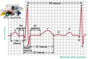
Each wave on the electrocardiogram represents a specific event in the heart’s electrical cycle. The P wave represents the electrical activity that causes the heart’s atria to contract, while the QRS complex represents the electrical impulses that cause the ventricles to contract. The T wave represents the recovery of the ventricles.
Why are electrocardiograms important?
Electrocardiograms are an invaluable tool in diagnosing and monitoring heart conditions. They provide valuable information about the heart’s electrical activity, allowing healthcare professionals to detect abnormalities and assess the overall health of the heart.
One of the primary uses of electrocardiograms is in diagnosing heart rhythm disorders, also known as arrhythmias. These are conditions in which the heart beats too fast, too slow, or irregularly.
By analyzing the waves on the electrocardiogram, healthcare professionals can identify the specific type of arrhythmia and determine the best course of treatment.
Electrocardiograms are also used to assess the risk of heart disease and other cardiovascular conditions. Certain wave patterns on the electrocardiogram can indicate the presence of underlying heart problems, such as coronary artery disease or an enlarged heart.
By identifying these patterns, healthcare professionals can initiate appropriate interventions to prevent further complications.
Additionally, electrocardiograms are often used to monitor patients who have already been diagnosed with heart conditions. Regular ECGs can help healthcare professionals track the progress of a condition, assess the effectiveness of treatment, and make any necessary adjustments to the patient’s care plan.
Common uses of electrocardiograms in healthcare
Electrocardiograms are used in a variety of healthcare settings and for various purposes. One of the most common uses is in the emergency department, where ECGs are routinely performed to assess patients with chest pain or other symptoms suggestive of a heart problem.
An ECG can quickly determine if a patient is experiencing a heart attack or another cardiac event, allowing for immediate intervention.
In addition to emergency situations, electrocardiograms are also performed in primary care settings as part of routine check-ups. They provide a baseline assessment of the heart’s electrical activity and can help identify any early signs of heart disease or other abnormalities.
Routine ECGs are particularly important for individuals with risk factors for heart disease, such as high blood pressure, diabetes, or a family history of heart problems.
Electrocardiograms are also used in specialized cardiac clinics and hospitals to assess patients with known heart conditions. They are often performed before and after cardiac procedures, such as angioplasty or heart surgery, to evaluate the heart’s response to treatment and ensure its proper functioning.
How to prepare for an ECG
Preparing for an electrocardiogram is generally a straightforward process that requires minimal effort from the patient. Here are a few simple steps to ensure a successful ECG procedure:
- Wear comfortable clothing: It is recommended to wear loose-fitting clothing that allows easy access to the chest area. Avoid wearing garments with metal buttons or zippers, as they may interfere with the electrode placement.
- Avoid applying lotions or oils: Before the ECG, it is important to refrain from applying any lotions, oils, or creams to the chest area. These substances can interfere with the electrode’s ability to adhere to the skin and may affect the accuracy of the test.
- Inform the healthcare professional of any medications: It is crucial to inform the healthcare professional conducting the ECG of any medications or supplements you are currently taking. Some medications, such as beta-blockers or calcium channel blockers, can affect the heart’s electrical activity and may influence the results.
- Relax and remain still during the procedure: During the ECG, it is important to remain as relaxed as possible. Movement or muscle tension can interfere with the quality of the recorded signals. The healthcare professional will instruct you on how to position your body to ensure accurate results.
By following these simple steps, you can help ensure a smooth and accurate ECG procedure.
What to expect during an ECG procedure
An electrocardiogram is a quick and painless procedure that typically takes around 5 to 10 minutes to complete. Here’s what you can expect during an ECG:
- Preparation: The healthcare professional will first explain the procedure and ask you to remove any clothing or jewelry that may interfere with the electrode placement. You will be provided with a gown or a sheet to cover yourself during the test.
- Electrode placement: The healthcare professional will attach small adhesive electrodes to specific locations on your chest, arms, and legs. These electrodes are connected to the ECG machine via wires. The number and placement of the electrodes may vary depending on the specific type of ECG being performed.
- Recording the ECG: Once the electrodes are in place, the healthcare professional will start the ECG recording by activating the machine. You will be asked to remain still and breathe normally during the recording process. The machine will produce a series of waves on its screen, indicating that the heart’s electrical activity is being recorded.
- Completion and removal of electrodes: After the recording is complete, the healthcare professional will remove the electrodes from your skin. The adhesive used is typically gentle and should not cause any discomfort. You may be provided with a towel or wipes to remove any residual adhesive.
- Interpretation of the ECG: The recorded ECG will be analyzed by a healthcare professional, such as a cardiologist or a trained technician. They will interpret the waves and patterns on the electrocardiogram and generate a report detailing their findings.
Interpreting an electrocardiogram
Interpreting an electrocardiogram requires knowledge and expertise in analyzing the various waveforms and patterns. While a detailed interpretation is best left to healthcare professionals, understanding the basic components of an ECG can help you gain insights into your heart’s health.

Here are some key elements to look for when interpreting an ECG [please refer to the above image for details]:
- Heart rate: The heart rate can be determined by calculating the number of QRS complexes per minute. A normal resting heart rate typically falls between 60 and 100 beats per minute. A heart rate above or below this range may indicate an underlying condition.
- Rhythm: The regularity of the waves and intervals on the ECG can provide important information about the heart’s rhythm. A regular, evenly spaced pattern is considered normal, while irregularities may indicate an arrhythmia or other heart rhythm disorder.
- P wave: The P wave represents the electrical activity that causes the atria to contract. It should be smooth and rounded, indicating normal atrial depolarization.
- PR interval: The PR interval represents the time it takes for the electrical signals to travel from the atria to the ventricles. A prolonged PR interval may suggest a conduction abnormality.
- QRS complex: The QRS complex represents the electrical impulses that cause the ventricles to contract. It should be narrow and consistent in shape, indicating normal ventricular depolarization.
- ST segment: The ST segment represents the interval between ventricular depolarization and repolarization. Deviations from the baseline may indicate myocardial ischemia or injury.
- T wave: The T wave represents the recovery of the ventricles. It should be upright and rounded, indicating normal ventricular repolarization.
Understanding these key components can provide a basic understanding of an ECG and help identify any potential abnormalities that may require further evaluation by a healthcare professional.
Common abnormalities seen in electrocardiograms
Electrocardiograms can detect various abnormalities and provide valuable insights into the heart’s health. Here are some common abnormalities that may be seen on an ECG:
- Arrhythmias: Arrhythmias are abnormal heart rhythms that can manifest as irregular patterns on the ECG. They can include conditions such as atrial fibrillation, ventricular tachycardia, or bradycardia. Learn more about Arrhythmia.
- Myocardial infarction: An ECG can show signs of a myocardial infarction, commonly known as a heart attack. This may include ST segment elevation or depression, T wave inversion, or the presence of Q waves.
- Conduction abnormalities: Conduction abnormalities can be detected on an ECG as prolonged PR intervals, bundle branch blocks, or other irregularities in the electrical conduction pathways of the heart.
- Hypertrophy: Electrocardiograms can indicate signs of cardiac hypertrophy, which is the thickening of the heart muscle. This may be seen as increased voltage in certain leads or other changes in the wave patterns.
- Ischemia: Ischemia, which refers to reduced blood flow to the heart muscle, can be detected on an ECG as ST segment changes or T wave inversions. These changes may indicate an increased risk of a future heart attack.
It is important to note that while ECGs can provide valuable information, they are not always definitive. Further evaluation and testing may be required to confirm a diagnosis or assess the severity of a condition.
Frequently asked questions about electrocardiograms
- Are electrocardiograms painful? No, electrocardiograms are completely painless. The electrodes are simply attached to the skin by leads, and there are no injections or invasive procedures involved.
- Can I eat or drink before an ECG? Yes, you can eat and drink as usual before an ECG. There are no dietary restrictions for this procedure.
- Who can perform an ECG? Electrocardiograms can be performed by trained healthcare professionals, such as nurses, medical technicians, or cardiologists.
- Can I exercise before an ECG? It is best to avoid intense physical exercise immediately before an ECG, as it can temporarily affect the heart’s electrical activity. However, light activities such as walking or climbing stairs should not interfere with the accuracy of the test.
- How long does it take to receive the results of an ECG? The time it takes to receive the results of an ECG may vary depending on the healthcare setting. In some cases, the results may be available immediately, while in others, it may take about 15 to 20 minutes subject to availabilty of the physician and for a healthcare professional to analyze and interpret the ECG.
- Can I drive after an ECG? Yes, you can drive after an ECG, unless you have been diagnosed with an heart attack that may need admission for observation and further care. The test does not have any sedative or impairing effects that would prevent you from operating a vehicle.
Conclusion: The importance of electrocardiograms in diagnosing and monitoring heart health
Electrocardiograms play a crucial role in diagnosing and monitoring heart conditions. They provide valuable insights into the heart’s electrical activity, allowing healthcare professionals to detect abnormalities, diagnose various heart conditions, and assess the overall health of the heart.
By understanding the basics of ECGs, including how they work, their uses in healthcare, and how to interpret the results, you can become an informed advocate for your own heart health. Whether you’re a healthcare professional wanting to refresh your knowledge or a curious individual seeking to learn more, this comprehensive guide has provided you with everything you need to know about electrocardiograms.
Useful References:
Final words:
I hope you have found this article useful. Please share and promote it with people you know using the icons at the bottom of this article. Alternately, if you have an X account [previuosly Twitter], please click to tweet below-
Electrocardiograms: Everything You Need to Know About It Share on X
