Have you ever been to a cardiologist recently and after clinical examination been told to get a 2D echocardiography done? Would you like to learn why you have been ordered this investigation?
Then look no further- you have come to the right place, because today we are going to learn about this unique investigation that tells us about our present heart health.

The human heart is one of the most enigmatic organs in the body, beating non-stop till you live. If it pauses or stops, then we wish- RIP to that person. It’s also one of the most crucial, as any doctor or your cardiologist will happily tell you.
Fortunately, this mysterious yet important organ has been thoroughly researched and studied over the years, so we know a lot about it.
However, researchers are still uncovering new insights into how it works and why some people develop problems with it.
One form of research that has recently gained traction is echocardiography (Echo). This test is non-invasive and uses special technology to analyze different aspects of cardiac output. In this blog post, we cover everything you need to know about Echocardiography and its benefits.
Table of Contents
What is echocardiography or ECHO?
ECHO is an emerging technology used to measure cardiac output. Cardiac output is a measurement of the volume of blood that the heart pumps out per minute. While this may not seem like something we should know, it is actually very important.
In fact, it’s used to indicate the general health of the heart and is a common indicator for heart disease and other cardiac problems. The amount of blood your heart pumps throughout the course of a few minutes is influenced by many factors, including your body size and the demands placed on your heart.
What does ECHO do for you?
The above short video shows how and what the cardiologist views while carrying out this procedure. ECHO allows doctors or researchers in cardiology to measure cardiac output without needing to draw blood by using an ultrasound. They use a special technology that passes light through the skin, allowing them to examine the flow of blood.
Echocardiography, also known as cardiac ultrasound, is a type of medical imaging that uses ultrasound to examine the heart. The visual image formed using this technique is called an echocardiogram, a cardiac echo, or simply an echo.
It is one of the most widely used diagnostic imaging modalities in cardiology and is routinely used in the diagnosis, management, and follow-up of patients with any suspected or known heart diseases.
Echocardiography can provide a wealth of helpful information, including the size and shape of the heart (internal chamber size quantification), pumping capacity, location and extent of any tissue damage, and assessment of valves.
An echocardiogram can also give physicians other estimates of heart function, such as
- a calculation of the cardiac output [explained below],
- ejection fraction, and
- diastolic function (how well the heart relaxes).
Echocardiography is an important tool in assessing heart wall motion abnormality in patients with suspected cardiac disease. It is a tool which helps in reaching an early diagnosis of myocardial infarction [heart attack], showing regional wall motion abnormality.
Also, it is important in treatment and follow-up in patients with heart failure, by assessing ejection fraction.
Echocardiography can help detect cardiomyopathies [disease or inflammation of heart muscle], such as hypertrophic cardiomyopathy and dilated cardiomyopathy.
The use of stress echocardiography may also help determine whether any chest pain or associated symptoms are related to heart disease.
The biggest advantage of echocardiography is that it is not invasive (does not involve breaking the skin or entering body cavities) and has no known risks or side effects.
Not only can an echocardiogram create ultrasound images of heart structures, but it can also produce accurate assessment of the blood flowing through the heart by Doppler echocardiography, using pulsed- or continuous-wave Doppler ultrasound.
This allows assessment of both normal and abnormal blood flow through the heart. Color Doppler, as well as spectral Doppler, is used to visualize any abnormal communications between the left and right sides of the heart, any leaking of blood through the valves (valvular regurgitation) and estimate how well the valves open (or do not open in the case of valvular stenosis).
The Doppler technique can also be used for tissue motion and velocity measurement by tissue Doppler echocardiography.
Echocardiography is performed by cardiac sonographers, cardiac physiologists (UK), or physicians trained in echocardiography.
What is Cardiac Output?
Cardiac output is the volume of blood that the heart pumps with each heartbeat. It is measured in litres per minute (L/min). While it’s an important metric, it’s not the only one used to measure cardiac health. Cardiac output is influenced by many factors, including blood pressure, the volume of blood in your body, sodium levels, and more.
Cardiac output is important because it’s a common indicator for heart disease and other problems with the heart.
Cardiac output can be measured in a variety of ways. It is commonly measured by drawing blood from the arm, which allows researchers to see the levels of oxygen and carbon dioxide in the blood. However, this is an invasive procedure that can be uncomfortable and cause bruising or other side effects.
How Does Echocardiography Work?

Echo, as we’ve mentioned, does not require blood to be drawn, as blood pressure and flow measurements can be made through the skin. This is a non-invasive procedure that uses light to measure the flow of blood through the skin and into the heart, lungs and brain.
The device uses a small amount of visible red light that is non-harmful and has no side effects. The light is passed through the skin and is absorbed by hemoglobin in the blood, which appears as a bright red color when it reaches the detector.
Since blood flows through your body in different directions, the device has to be rotated to get a full picture of the blood flow.
Most devices have a built-in motor that can rotate the light source. This information is then processed to calculate the volume of blood flowing through the body in different directions.
Types of Echocardiography
There are several types of echocardiography that can be used to diagnose and monitor heart conditions. Here are some of the most common types:
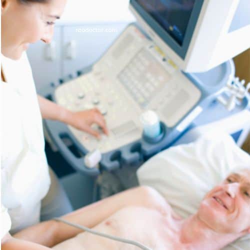
- Transthoracic echocardiogram (TTE): This is the most common type of echocardiogram and is noninvasive, taking place entirely outside your body. A team member applies gel to your chest, then uses a handheld transducer to scan your heart. This test can provide information about the size and shape of your heart, how well your heart is pumping, and whether there are any problems with your heart valves.
- Transesophageal echocardiogram (TEE): This type of echocardiogram involves guiding a special ultrasound probe into your mouth and down your esophagus after sedation. This approach provides better images than TTE because the esophagus and heart sit close together, and the sound waves do not need to pass through skin, muscle, or bone. TEE is a better choice for some conditions, such as when doctors need to see a specific part of the heart with greater resolution.
- Stress echocardiogram: This test is done as part of a stress test that deliberately increases your heart rate and blood pressure. We take two sets of images, one at rest and another after working out on a treadmill or stationary bike. If your health prevents such physical activity, we inject a medication that mimics the effect of exercise.
- Doppler echocardiogram: Doppler echocardiography is used to measure and assess the flow of blood through the heart’s chambers and valves.
- Color Doppler: This technique is used to visualize any abnormal communications between the left and right sides of the heart, any leaking of blood through the valves (valvular regurgitation) and estimate how well the valves open (or do not open in the case of valvular stenosis).
- M-mode echocardiography: This is the simplest type of echocardiography that produces an image similar to a tracing rather than an actual picture of heart structures. M-mode echo is useful for measuring heart structures, such as the heart’s pumping chambers, the size of the heart itself, and the thickness of the heart walls.
- Intracardiac echocardiography (ICE): This newer form of testing involves taking images inside your heart using a catheter inserted through a vein in your groin or neck.
Steps in performing Echocardiography-how echocardiogram done-

As mentioned above, the Transthoracic method is the most commonly used method. this procedure involves some steps as given below:
- The patient is asked to lie down on an examination table and the cardiologist/technician applies gel to the chest area.
- He then places a transducer, which is a small handheld device, on different areas of the chest to obtain images of the heart.
- The transducer emits high-frequency sound waves that bounce off the structures of the heart and create images on a monitor.
- The technician moves the transducer around to capture images of different angles of the heart.
- The patient may be asked to hold their breath or change positions during the procedure to get clearer images.
- The images obtained are recorded and reviewed by a cardiologist or a specialized technician to assess the structure and function of the heart.
- In some cases, a stress echocardiogram may be done, where the patient is asked to exercise on a treadmill or receive medication to make the heart work harder, while echocardiogram images are taken simultaneously.
- The procedure usually takes around 30-60 minutes and is painless.
This example will show how it is done:
- The patient arrives at the cardiology clinic and is greeted by the cardiologist or ECHO technician.
- He/she then explains the procedure to the patient and asks him/her to remove their clothing from the waist up and put on a gown.
- The patient is then led to the examination room and asked to lie down on the examination table.
- The doctor/technician applies a clear gel to the chest area, which helps with the transmission of sound waves.
- The technician then places the transducer on different areas of the chest, such as the upper chest, near the breastbone, and under the left breast, to obtain images of the heart.
- The patient may be asked to hold their breath or change positions during the procedure to get clearer images.
- The technician moves the transducer around to capture images of different angles and structures of the heart.
- The patient may feel slight pressure or discomfort during the procedure, but it should not be painful.
- The images obtained are recorded and reviewed by a cardiologist or a specialized technician to assess the structure and function of the heart.
- The patient is then allowed to clean off the gel and get dressed.
- The cardiologist or technician will discuss the results of the echocardiogram with the patient during his/her consultation.
Useful reference: Stanford Healthcare
Benefits of Echocardiography
While many medical tests have their advantages and disadvantages, eco-cardiography has several benefits that set it apart.
Many of these are the result of the non-invasive nature of the test. As we’ve mentioned, it doesn’t require blood to be drawn, which means it doesn’t risk compromising blood flow in other parts of the body. This can be advantageous for people with weak or damaged veins that are difficult to access.
Because no blood needs to be drawn, it also doesn’t need to be sterilized or disposed of, which can be costly and time-consuming. Additionally, it has been shown to be more accurate than blood tests for people of all ages and health conditions.
This is partially due to the fact that it doesn’t require any calibration and can make adjustments based on the individual being tested.
Limitations of ECHO
While eco-cardiography is non-invasive, it still requires that a device be placed near the skin to measure blood flow.
This means that certain conditions may make it difficult to use. People with wounds that may be irritated by the device or those who have sensitive skin may not be good candidates.
In addition, people with very dry skin or who have applied lotion or other products near the skin may not get accurate readings.
Key Takeaway
Echocardiography is an emerging technology used to measure cardiac output. Cardiac output is a measurement of the volume of blood that the heart pumps out per minute.
The device uses a small amount of visible red light to measure blood flow through the skin, allowing it to be used without drawing blood or using an ultrasound.
It has several benefits that other tests don’t, including the fact that it is non-invasive and more accurate than blood tests. Its limitations include the need to have the device near the skin and that certain conditions may make it difficult to use.
Useful References:
Final Words
I hope you have found this article useful. If yes, please share within your friend and family circle using the share buttons at the bottom of the article. Alternately, Click to Tweet below-
ECHOCARDIOGRAPHY: What You Should Know and Why It Matters Share on XMy next article will be on a very common, yet distressing ailment experienced nowadays- Dyspepsia or Hyperacidity.


